LEPROSY (Hansen's disease) / MedUrgent
LEPROSY (Hansen's disease)
Armauer Hansen in Bergen (Norway) in 1873 descried Mycobacterium leprae as the first reported bacterial pathogen in humans in a fresh mount of scraping from a leproma of a Norwegian leprosy patient. M.Leprae is an acid-fast bacillus that does not grow on the routine mycobacteriologic media. It multiplies slowly and when killed leads to strong reactions as in tuberculoid leprosy patients while lepromatous patients do not show this reaction. It is the most common communicable disease affecting the peripheral system and the skin world-wide, but now it is mainly confined to the developing world.
Pathogenesis:
In Sudan, it is endemic in the South, Nuba Mountains and Central Sudan. The mode of transmission is not uniform and skin-to-skin contact, nasal secretions, breast feeding, flies, sexual intercourse and blood transfusion and prolonged contact by a susceptible subject with a source, as in the case of husband and wife and children are all possible routes. Naturally acquired leprosy has been described in chimpanzee and other monkeys, a reason to believe that leprosy may be related to zoonosis. M.leprae in tissues generates a reaction involving lymphocytes, multinucleated giant cells, epithelioid cells and histiocytes forming a granuloma. It destroys Schwann cells of nerves leading to loss of sensation. The sweat glands, hair follicles and melanocytes are all destroyed leading to lack of sweat, loss of hair and hypopigmentation.
• Lepromatous
Leprosy: There is depressed cellular immunity and histiocytes are large in size
and have a foamy appearance with a large number of bacilli included (Lepra or
Globi cells). This effect involves skin, liver, spleen, lymphocytes, bone
marrow, testes, bone and kidneys.
• Borderline and Intermediate forms of leprosy
may show similar reactions but to a lesser degree. In all types of leprosy,
systemic manifestations of toxicity e.g. fever, anemia and loss of weight are
minimal.
• Tuberculoid leprosy the inflammatory process starts in the peripheral nerves extending to varying depth in the subcutaneous tissue along the nerve fibers. This is followed by bacillary invasion of Schwann cells and swelling of the peripheral nerves from where it is transmitted to the skin.
CLINCAL PICTURE:
The incubation period varies from a few months to 30 years. Onset of the disease is usually gradual. An area of impaired sensation without systemic manifestations is an early sign of the disease. It is uncommon among children but may occur due to close contact with affected parents particularly at night. Clinical presentations include:
Indeterminate Leprosy
This
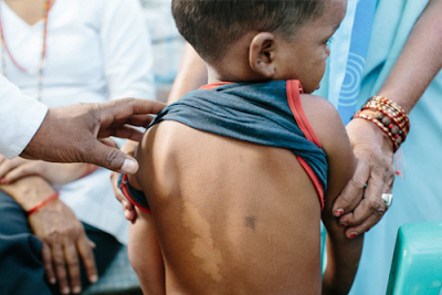
type presents with a single or more poorly defined macules that may heal
spontaneously or progress to Tuberculoid Leprosy (TT), or Lepromatous Leprosy
(LL). Effect on color, sensation and sweating is minimal and M. Leprae may not
be isolated from skin smears but histopathology is diagnostic. This type is
most commonly seen in children age 2 -15 years.
Tuberculoid Leprosy
A few
well demarcated hypopigmented macules develop de novo or secondary to
indeterminate leprosy. The size of macules varies and there is impaired
sensation, loss of hair and sweating. Adjacent nerve trunks are palpable and
tender. Unlike the lepromatous form, it occurs late, is asymmetrical and is
localized to one or two nerves. The skin becomes thickened and loss of the
sensations is more common than loss of the motor functions. The 5th cranial
nerve involvement leads to loss of sensation on the face while peripheral nerve
involvement leads to loss of sensation in the legs and soles of feet.
Tuberculoid leprosy tends to heal spontaneously but slowly, often without any
residual disability.
Lepromatous Leprosy
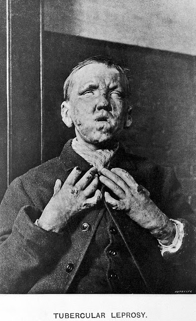
This is a widely disseminated form of leprosy. Juvenile form of LL is difficult to detect since the macules have indistinct boarders and there are no alterations in sensation or sweating and Acid Fast Bacilli are only few in these lesions. Histopathology is the only diagnostic tool. The disease usually progress to form plaques, papules and nodules. Macules are, hypopigmented lesions most commonly seen on the face, limbs and buttocks. Papules and Nodules are advanced lesions that involve ears, nose, face, palms and buttocks. Thickening of the skin with the appearance of papules and macules and lymphoedema gives the appearance of 'Leonine face'. Lepromatous Leprosy leads to loss of hair of the scalp and face (eye brows, eye lashes and beard), destruction of the nasal bone and mucus membranes (saddle nose), keratitis and lagophthalmia (failure of lower eye lid to close). Nerve are affected late leading to sensory loss, loss of temperature sensation, pin prick and touch sensation. Affection of motor nerves leads to claw hands, drop feet and facial palsy. Autonomic nervous system may are be involved in advanced cases. Bone changes include periostitis, bony cysts, aseptic necrosis and osteomyelitis. This could then be followed by the middle and proximal phalanges destruction. Acute amputation caused by peripheral neuropathy, CNS involvement, trauma and bone infection. Joint involvement leads to the formation of "Charcot's joints".
CLASSIFICATION
Leprosy has been classified according to the
clinical, pathological and immunological pictures at the time of presentation of
the patient. Two stable forms are known. These are tuberculoid leprosy (TT) and
lepromatous leprosy (LL) which are determined by the level of cell-mediated
immunity of the individual. Those with poor immunity develop LL, while those with good immunity develop TT. Intermediate immunity leads to borderline or
dimorphous picture. This is an unstable form that may, if untreated, progress
to either LL or TT. In between there are two varibles BT and BL, that show
features of both ends of the spectrum.
Lepra Reactions
Two types of acute reaction episodes are known to occur during the course if the disease, with or without treatment.
1- Reversal Reaction
This is a delayed hypersensitivity reaction seen in Borderline leprosy. The lesions become erythematous, edematous and tender with acute neuritis. The condition may progress to TT or heal spontaneously. This form is not seen in LL.
2- Erythema Nodosum Leprosum
This is seen in
LL and BL after a few months of starting the treatment. presents as erythematous tender
subcutaneous nodules, fever, synovitis and iridocyclitis. The reaction frequently
leads to extensive ulceration of the skin. This may end up with
glomerulonephritis and amyloidosis Management of both types of reactions includes
aspirin, corticosteroid and thalidomide in severe cases.
INVESTIGATIONS
- Skin snip: staining with ZN for acid – fast
bacilli. The areas chosen are elbows, ear lobes or wherever the lesions are
obvious.
- Skin biopsy for histopathology: This is
rather useful for the tuberculoid form
- Nerve biopsy: This is performed when all
other tests have proved negative.
The biopsy is taken from superficial nerves
such as the Great Auricular Brachial and Radial nerves.
- Bacteriological examination: Take a nasal
smear and stain it by a ZN stain, bacteria can be seen in LL but not usually in
the other forms
- Serology
- Hypergammaglobulinemia in LL
- Lepromin Test: Intradermal inoculation of
killed M.Leprae. It is of no diagnostic value, but a positive reaction in
treated cases of LL is a good prognostic sign as it indicates good cellular
immunity. It is usually positive in normal people, TL, and BL. It is negative
in untreated LL.
TREATMENT
Gain the patient's confidence to understand and scope of the disease, its stigmata, side-effects of drugs and how to look after the anesthetic limbs and acceptance of the long duration of treatment.

* Treatment should continue for 5-10 years or
until 3 consecutive skin smears are negative for bacilli
Treatment in children
• Dapsone 10 – 25 mg/day
• Clofazimine 200 - 300 mg/day in 2-3 divided
doses
• Rifampicin 10 mg/kg/day Prevention
prevention
• Health education
• Decrease contact with positive cases
• Care of ulcers, hands and feet
• Social and psychological support
• BCG vaccine to enhance cellular immunity
against mycobacterium.


.JPG)


Comments
Post a Comment